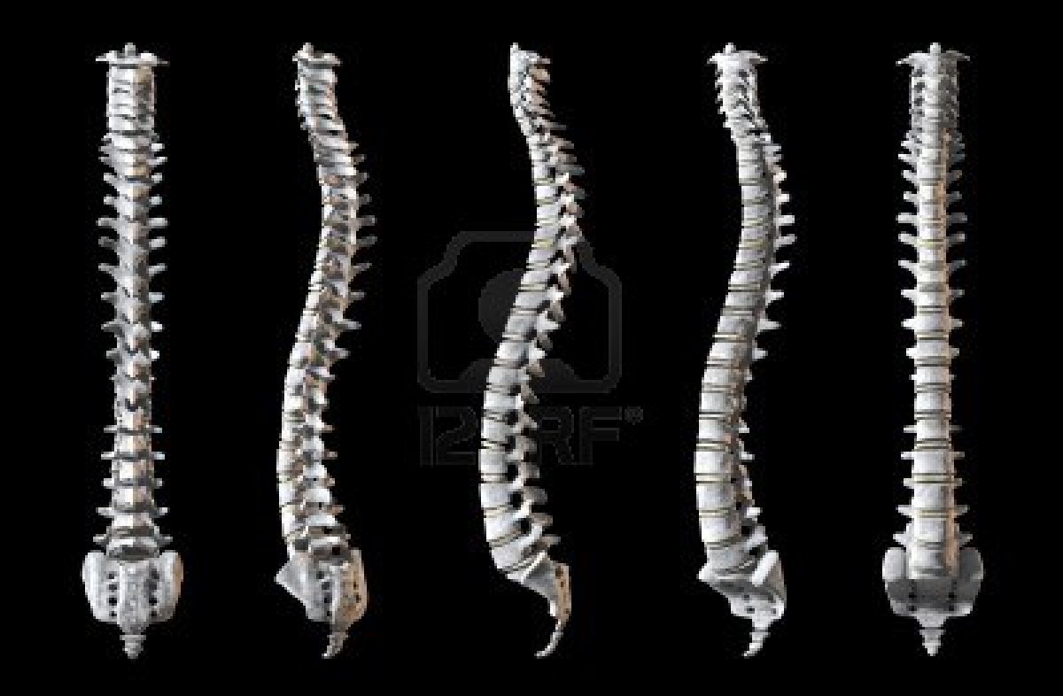
The human spine is made up of 33 vertebrae, but how do they support our bodies while allowing us such flexibility?
The human spine is made up of 33 vertebrae, 24 of which are articulated (flexible) and nine of which normally become fused in maturity. They are situated between the base of the skull to the pelvis, where the spine trails off into the coccyx – an evolutionary remnant of a tail our ancestors would have displayed.
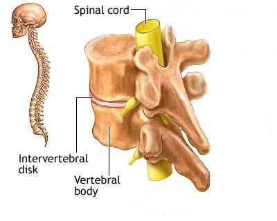
The primary functions of the vertebrae that make up the spine are to support the torso and head, which protect vital nerves and the spinal cord and allow the individual to move. By sitting closely together, separated only by thin intervertebral discs which work as ligaments and effectively form joints between the bones, the vertebrae form a strong pillar structure which holds the head up and allows for the body to remain upright. It also produces a base for ribs to attach to and to protect vital internal organs in the human body.
Vertebrae are not all fused together because of the need to move, and the vertebrae themselves are grouped into five types – cervical, thoracic, lumbar, sacral and coccygeal. The sacral vertebrae fuse during maturity (childhood and teenage years) and become solid bones towards the base of the spine. The coccygeal vertebrae will fuse in some cases, but studies have shown that often they actually remain separate. Collectively they are referred to as the coccyx (tail bone).
Spinal cords and nerves
Spinal cord injuries are normally caused by trauma. If the trauma causes intervertebral discs and vertebrae to break, they can pierce the spinal cord, which can result in loss of feeling. Cord severance may result in paralysis.
Spine curvature
As you look at the human spine, you can see some distinct curves. The primary reasons for these are to help distribute weight throughout the spine and support aspects of the body. The curve most familiar to us is the lumbar curve, between the ribs and pelvis. This develops when we start to walk at about 12-18 months and helps with weight distribution during locomotion. Prior to this we develop the cervical curve, which allows us to support the weight of our head at around three to four months, and two smaller less-obvious curves in the spine (the thoracic and pelvic curves) are developed during gestation.
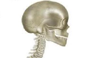
The skull is connected to the spine by the atlanto-occipital joint, which is created by CI (atlas) and the occipital bone situated at the base of the cranium (skull).
This unique vertebra has no ‘body’ and actually looks more like a ring than any other vertebra. It sits at the top of the cervical vertebrae and connects with the occipital bone via an ellipsoidal joint, allowing movement such as nodding or rotation of the head. An ellipsoidal joint is where an ovoid connection (in this case the occipital bone) is placed into an elliptical cavity (CI vertebrae). The rest of the cervical vertebrae also work to support the weight of the head.